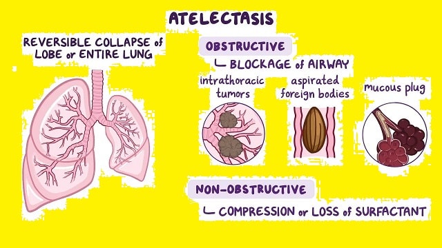
Atelectasis
Atelectasis is a condition that occurs because the part called the alveoli in the lungs is not filled with air. As a result, the lungs fail or do not expand perfectly. Normally, effective breathing takes place from the time the nose inhales air, until the air enters the respiratory tract, namely the bronchi, and bronchioles, and reaches the alveoli. Then from the alveoli, air will enter the blood vessels and the oxygen content of the air will be distributed throughout the body.
In order to function normally, the alveoli need to always be in an open state, so that air containing oxygen can enter and flow into the blood vessels. This condition is influenced by several things, such as surfactant fluid (to maintain surface tension), continuous breathing activity (to keep the alveoli open), deep breathing (to encourage the release of surfactant into the alveoli ), and coughing (to clear the respiratory tract of mucus).
Atelectasis is not actually a disease, but a radiological picture caused by a disease. If left untreated, this condition can lead to a lack of oxygen in the body and infections, such as pneumonia.
Symptoms of Atelectasis
Complaints that may be experienced by atelectasis sufferers can vary, from no complaints at all to severe complaints depending on the number of alveoli affected. The more alveoli affected, the greater the likelihood of symptoms appearing, such as:
- Hard to breathe.
- Chest pain, especially when breathing deeply or coughing.
- Breathing becomes rapid.
- Heart rate increases.
- The skin on the body, lips, fingertips, and toes turns bluish.
If atelectasis is accompanied by a lung infection, additional symptoms may occur in the form of a productive cough and fever.
Causes of Atelectasis
Atelectasis can be caused by many things. Based on the cause, this condition can be divided into five, namely obstructive, passive, compressive, adhesive, and cicatricial atelectasis.
- Obstructive Atelectasis
This type is the most common type of atelectasis. The cause can be due to a blockage, either inside or outside the airway, which interferes with the development of the alveoli. Blockages can be caused by foreign objects, tumors, thick secretions, and infections.
- Passive Atelectasis
This type of atelectasis is also called relaxation atelectasis. This condition is caused by the presence of a mass outside the lung that inhibits lung expansion. This mass can be a buildup of fluid in the pleura (for example, blood, transudate, or exudate ) or air ( pneumothorax ) that causes the lung to collapse. In addition, elevation of the diaphragm or herniation of abdominal organs can also press on the lungs.
- Compressive Atelectasis
This atelectasis is similar to passive atelectasis, but the mass is located within the lung. The cause can be a large bulla, malignancy, abscess, or other lesion that causes compression of the lung.
- Adhesive Atelectasis
Adhesive atelectasis is a type of non-obstructive and non-compressive atelectasis due to reduced surfactant production by type II pneumocytes. The causes of pneumocyte damage can vary, including genetic disorders, general anesthesia, ischemia, and radiation damage. This type is rare and is usually found in children with hyaline membrane disease. In addition, adhesive atelectasis can also be caused by acute respiratory distress syndrome, smoke inhalation, heart bypass surgery, uremia, and prolonged shallow breathing.
- Cicatricial Atelectasis
This type of atelectasis describes the scarring and contracture of the lung tissue that occurs after lung infection (eg, pneumoconiosis ), scleroderma, radiation, and idiopathic pulmonary fibrosis. This condition can occur locally, for example, with tuberculous infection of the upper part of the lung, or generally, for example, with interstitial pulmonary fibrosis.
Risk Factors for Atelectasis to Watch Out For
Atelectasis can happen to anyone regardless of gender or race. However, there are several factors that can increase the risk of this disease, including:
- Aged 60 years or older.
- The baby was born prematurely.
- Completed surgery.
- Have lung disease.
Diagnosis of Atelectasis
Examinations that can be performed to diagnose atelectasis include:
- Examination of medical history and complaints.
- Physical examination.
- Supporting examinations, such as:
- Blood oxygen level check using an oximeter. This tool will be attached to the fingertip.
- Examination of oxygen, carbon dioxide, and blood chemistry levels with blood gas examination.
- Chest X-ray. This examination can see the general condition of atelectasis.
- CT Scan Examination. Through this examination, possible causes of atelectasis, such as tumors or other masses can be identified.
- Bronchoscopy examination. This examination is to see if there is a blockage in the airway. The doctor will insert a camera using a special tool through the nose or mouth, down to the lungs.
Atelectasis Treatment
Treatment for atelectasis is given based on the cause. In general, treatment for this condition can be done with non-surgical therapy and surgical therapy.
- Non-surgical Therapy
Most cases of atelectasis can be treated with:
- Chest physiotherapy. This therapy is done by moving the body to help smooth the airways. This therapy is often used to cure people with obstructive atelectasis, post-surgery, and cystic fibrosis.
- Bronchoscopy. This examination is done by inserting a camera and special tools into the airway to remove blockages.
- Breathing exercises. Breathing exercises can be done with the help of physiotherapy or with special tools, such as an incentive spirometer to help breathe deeply and open the alveoli. Usually, this therapy is done in patients with post-surgical atelectasis.
- Drainage. This procedure is performed when atelectasis is caused by pneumothorax or pleural effusion. In the drainage process, the doctor will insert a special needle and attach a special tube to drain air, fluid, or blood into a special bag.
- Administration of Antibiotics and Other Medications. This action is given according to the cause of atelectasis.
- Surgical Therapy
In certain conditions, it may be necessary to remove part of the lung tissue. For this, surgery is needed.
Prevention of Atelectasis
Prevention of atelectasis can be done by implementing a healthy lifestyle, including:
- Eat healthy foods.
- Drink enough water.
- Regular exercise.
- Avoid smoking.
- Avoid inhaling excessive smoke.
- Have regular treatment with a doctor if you have lung disease.
If you have lung problems and are planning to have lung surgery, you should discuss this with your doctor to avoid atelectasis after surgery.
Prevention of this disease can also be done if the immune system is strong enough. Therefore, complete a healthy lifestyle by consuming additional multivitamins so that the immune system is maintained strong.

