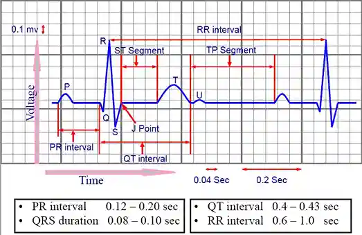
What is an EKG (Electrocardiogram)?
An electrocardiogram or ECG is a non-invasive diagnostic tool that records the electrical activity of the heart through electrodes placed on the surface of the body.
The ECG serves to visualize the electrical patterns of the heart produced during depolarization and repolarization of the myocardium, which are then interpreted to assess the electrical and mechanical function of the heart.
The ECG is an important tool for diagnosing a variety of cardiovascular conditions, including arrhythmias, myocardial ischemia, myocardial infarction, electrolyte disturbances, and drug effects on the heart.
ECGs are also used to monitor heart health routinely or as part of a clinical evaluation in patients with suspicious symptoms.
This procedure is considered safe, fast, and painless because it is performed without electric current and without incisions (non-invasive).
Types of ECG
According to the book Basic and Bedside Electrocardiography by Romulo F. Baltazar, ECG has several main types that are used according to clinical needs and patient conditions.
The following are the types of ECG described in the book:
-
Standard 12-Lead Electrocardiogram
Standard 12-lead EKG is the most common type that uses 12 leads (electrodes) to record the heart’s electrical activity from multiple angles.
Used to diagnose arrhythmias, ischemia, myocardial infarction, and cardiac hypertrophy.
-
Single-Lead Electrocardiogram (Rhythm Strip)
Uses one lead (usually lead II or V1) to continuously record the heart rhythm.
Useful for monitoring arrhythmias or short-term changes in heart rhythm.
-
Holter Monitoring
Continuously records ECG for 24-48 hours or more using a portable device.
Used to detect episodic arrhythmias, silent occult ischemia, or inconsistent symptoms.
-
Event Recorder
Similar to a Holter, but only records when the patient presses a button during symptoms.
Suitable for patients with rare arrhythmia symptoms.
-
ECG Stress Test (Treadmill Test)
Recording an ECG while the patient is exercising to evaluate myocardial ischemia or stress tolerance.
Useful in diagnosing coronary artery disease.
-
Signal-Averaged ECG (SAEKG)
Using computerized techniques to detect small electrical potentials that may indicate the risk of ventricular arrhythmia.
Usually used in post-myocardial infarction patients.
-
Ambulatory Blood Pressure Monitoring with ECG
A combination of periodic blood pressure monitoring and ECG recording to evaluate the relationship between blood pressure and the electrical activity of the heart.
-
Intraoperative ECG Monitoring
Performed during surgery to monitor heart conditions in real-time, especially during anesthesia.
Focus on detecting arrhythmias or sudden ischemia.
These types of ECG are selected based on clinical indications, duration of symptoms, and diagnostic purposes.
Electrocardiogram Function
In general, an electrocardiogram functions to determine how the heart works and detect any disorders in the organ.
Some heart diseases that can be detected through an electrocardiogram include:
- Cardiomyopathy or disorders of the heart muscle
- Arrhythmia or irregular heartbeat
- Heart attack
- Coronary heart disease
In addition, an electrocardiogram can also detect other disorders of the heart, such as electrolyte imbalances or side effects of drugs that affect heart rhythm.
Why Do an Electrocardiogram (ECG)?
An electrocardiogram is performed if you experience symptoms of heart disease, such as chest pain, difficulty breathing, fatigue, weakness, palpitations, and heart rhythm disturbances (tachycardia or bradycardia).
This test aims to detect health problems related to the heart, such as heart attacks, coronary heart disease, electrolyte disorders, poisoning, and drug side effects, as well as evaluating the effectiveness of the pacemaker used.
Side effects of ECG include allergic reactions on the skin due to electrodes attached to the body.
Intermittent heart abnormalities are sometimes difficult to detect with just an ECG examination.
In this case, the heart abnormality is detected by examining the heart’s electrical activity which is slightly different from a standard ECG examination, namely:
- Stress test or treadmill ECG test. This is a non-invasive test to assess the heart’s response to physical activity. The patient walks or runs on a treadmill at increasing intensity, while the ECG, blood pressure, and symptoms are monitored to detect myocardial ischemia or heart rhythm disturbances.
- Holter monitor. This is a non-invasive test that continuously records the electrical activity of the heart for 24-48 hours or more. The patient wears a portable device to detect arrhythmias, ischemia, or other heart problems that may not show up on a standard ECG.
What Does Research Say?
According to a study in the journal Hearts in 2021, an electrocardiogram (ECG) not only records the electrical activity of the heart, but is also able to detect various conditions such as arrhythmia, heart disease, electrolyte disturbances, and lung disease.
Although the ECG is a simple, rapid, non-invasive, and cost-effective diagnostic tool, its effectiveness depends largely on the accuracy of its interpretation.
However, with the increasing use of computer analysis for ECGs, the ability of medical personnel to read ECGs manually has begun to decline.
This study also reviews the potential of artificial intelligence-based ECG algorithms (AI-ECG) that can help interpret, diagnose, and stratify patient risk more efficiently.
The findings are expected to improve the quality of patient care and support clinical workflows.
Fun Facts
1. EKG was first discovered in 1903 by Willem Einthoven.
2. The EKG procedure only takes about 5-8 minutes and is completely painless.
When to Do an Electrocardiogram (ECG)?
You need to have an EKG if you are at risk of heart disease because there is a history of heart disease in your family.
Other conditions that require an EKG examination are active smokers, people with obesity, diabetes, high cholesterol, or high blood pressure.
An EKG examination is also needed if you experience the following symptoms:
- Chest pain.
- Hard to breathe.
- Dizzy.
- Sudden fainting.
- Fast or irregular heartbeat (palpitations).
An EKG is often performed to monitor the health of people who have been diagnosed with heart problems, to help evaluate an artificial pacemaker, or to monitor the effects of certain medications on the heart.
Difference between Electrocardiogram and Electroencephalogram
Although their names sound similar, electrocardiograms and electroencephalograms are two different things.
An EKG is an examination performed to analyze the heart organ, while an electroencephalogram is a medical test performed to check activity in the brain.
However, both have something in common, namely monitoring the electrical impulses produced by the organ in question.
That is the explanation of the electrocardiogram. This examination is an important medical procedure for sufferers with heart complaints.
How is an Electrocardiogram (ECG) Performed?
So, here is the procedure for performing an EKG:
-
Before Electrocardiogram (ECG)
There is no special preparation for an EKG examination because this examination is generally performed in emergency situations.
An ECG is performed to detect heart attacks and to determine heart conditions that accompany other diseases.
If this examination is planned, you are advised to avoid using lotions, oils, or powders on your body (especially the chest area).
This can make it difficult to attach electrodes to the body. Make sure the doctor knows the history of medication and supplements that have been and are being consumed.
-
Implementation of Electrocardiogram (ECG)
The EKG is short, only about 5–8 minutes. You will be asked to remove your top and any accessories in your pockets before the exam.
During the examination, electrodes are attached to the chest, arms, and legs.
The electrodes installed are usually 10 or 12 in number, made of plastic, and small in size.
Each electrode wire is connected to an EKG machine to record the heart’s electrical activity.
The doctor will interpret the examination results on the monitor screen, then the results are printed on paper.
-
After Electrocardiogram (ECG)
You can resume your normal activities after an ECG is performed unless the results are abnormal.
In this case, certain activities will be limited according to the disease suffered.
The results of the EKG recording can be discussed with your doctor, or you can make an appointment to discuss them again.
Information that can be obtained from an ECG examination includes heart rate, heart rhythm, changes in the structure of the heart muscle, and oxygen supply to the heart.
Further examinations were carried out according to the findings of the ECG.
Where is an Electrocardiogram (ECG) Performed?
An EKG examination can be performed at a healthcare provider or clinic that provides an EKG machine.
Frequently Asked Questions (FAQ)
-
How is the EKG installation process?
The following are the stages of installing an EKG device:
- Preparation: The patient will be asked to lie on their back on a bed or stretcher. Some areas of skin on the patient’s chest, arms, and legs may need to be cleaned or shaved to ensure the EKG electrodes adhere properly.
- Electrode placement: EKG electrodes are small sensors that are attached to the patient’s skin. Typically, there are 12 electrodes placed on different areas of the body, including the chest, arms, and legs. Each electrode is connected to the EKG machine by wires.
-
How to read an ECG?
Reading EKG results requires an understanding of the waves, intervals, and segments displayed on the EKG graph. EKGs can be read by a doctor or trained health care professional.
-
What is a cardiac ECG?
A cardiac ECG is a medical test used to record the electrical activity of the heart. This test is very important in diagnosing and monitoring various heart conditions. A normal ECG shows a healthy heart that is beating at a regular rhythm.

